Using the Bilateral Tab
In the Bilateral workspace tab, you can examine specific regions of interest in depth, specifically the temporomandibular joint (TMJ) or ear. The view screens that appear on this workspace tab will depend on the type of acquisition you use. If you acquire only one side of the volume, then only the view screens relevant to that side are displayed.
Using the Horizontal Toolbar in Bilateral
Like MPR and Curve, you can create annotations, shapes, screenshots, reports, and export functions within Bilateral.
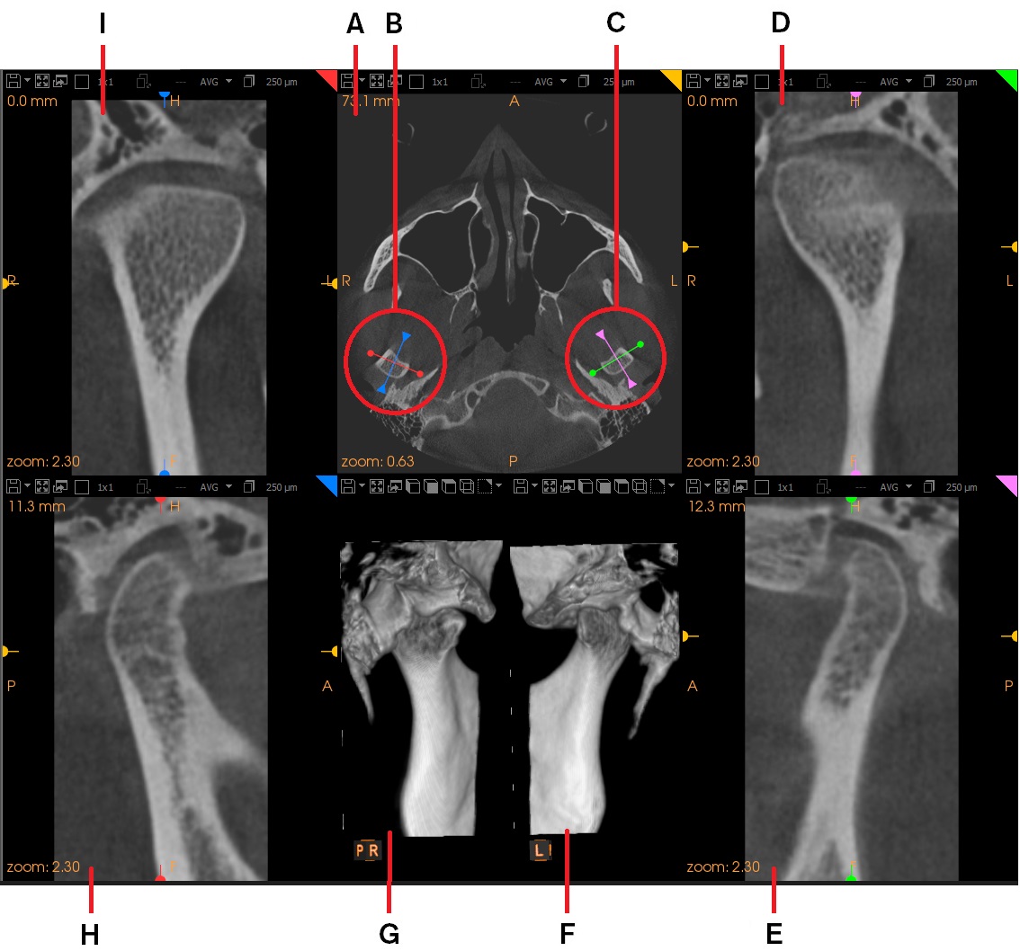
By default, the TMJ/Ear View Screen, TMJ/Ear Cross-Section View Screen and 3D View Screen are displayed. If the field of view is large enough, a second set of these view screens is displayed for the other side of the head.

The Bilateral workspace tab can therefore display either four or seven view screens. The following example shows all seven view screens.
|
|
View Screens |
Description |
|
A |
Axial Slice View Screen
|
This view screen shows a horizontal slice through the volume. In this view screen, you can draw a TMJ/ear trace over a region of interest on one side of the volume. If the acquired volume is large enough, the software automatically draws a mirror image trace on the opposite side of the volume. The colors in the handles in this view screen (B, C) match with the traces in the corresponding cross-section view screens. Once these traces have been drawn, the TMJ/Ear View Screen and TMJ/Ear Cross-Section View Screen appear, displaying slice views through the volume at the location of the traces. A 3D View Screen displays cropped TMJ or ear images. |
|
D |
LEFT TMJ/Ear View Screen |
This view screen appears when you draw a trace on the Axial Slice View Screen. In the Axial Slice View Screen, the trace is shown as a colored line. To move this slice plane, click and drag |
|
E |
LEFT TMJ/Ear Cross-Section View Screen
|
This view screen appears when you draw a trace on the Axial Slice View Screen. A view at 90° to the RIGHT TMJ/ear trace drawn on the axial slice is displayed. In the Axial Slice View Screen, the trace is shown as a colored line. To move this slice plane, click and drag |
|
F |
LEFT 3D View Screen (E)
|
Before any traces are drawn, this view screen and the RIGHT 3D View Screen (E) display identical views of the full volume. When you draw traces on the Axial Slice View Screen (A), the 3D View Screen displays the 3D view regions defined by the TMJ/ear cross-section and TMJ/ear cross-section traces. |
|
G |
RIGHT 3D View Screen (F) |
Before any traces are drawn, this view screen and LEFT 3D View Screen (D) display identical views of the full volume. When you draw traces on the Axial Slice View Screen (A), then the 3D View Screen displays 3D views of the regions defined by the TMJ/ear and TMJ/ear cross-section traces. |
|
H |
RIGHT TMJ/Ear Cross-Section View Screen |
This view screen appears when you draw a trace on the Axial Slice View Screen. A view at 90° to the LEFT TMJ/ear trace drawn on the axial slice is displayed. In the Axial Slice View Screen, the trace is shown as a colored line. To move this slice plane, click and drag |
|
I |
RIGHT TMJ/Ear View Screen |
This view screen appears when you draw a trace on the Axial Slice View Screen. In the Axial Slice View Screen, the trace is shown as a colored line. To move this slice plane, click and drag |
|
|
Note:
|
 in the
  in the
  in the
  in the